 Now that your cat’s teeth are sparkling it is important to begin a dental care regime to prevent them from getting into the same condition again. We understand that this can be difficult, so below are some tricks and information which should help. Continue reading
Tag Archives: dental
Part 2: My Cat needs dentistry surgery – But what does that actually involve?
12:15pm
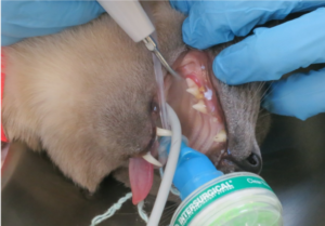
 Now that all of the teeth have been descaled, the vet is able to fully assess how healthy each one is. At the same time, they can examine the gums and mouth, looking for signs that indicate dental and/or oral disease. The tartar often covers the tooth so it can’t be examined until this stage. The vet will use a periodontal probe to examine each tooth, checking probing depth (the depth of the area between tooth and gum – a gap, or pocket of more than 1mm indicates that the tooth may need to be removed). They are also looking for resorption lesions, areas of infection, tooth mobility, furcation exposure (when the area between the roots of multi-rooted teeth is exposed due to bone loss), fractured or missing teeth and periodontal disease (gingivitis and periodontitis).
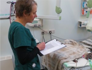
The Vet then records all of their findings on a dental chart. This is a crucial component of your cat’s medical history, and will be kept with their personal file for life.
12:20pm
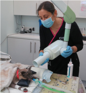 The next step is dental radiographs (x-rays). Cambridge Cat Clinic uses state-of-the-art digital dental radiography which provides us with clear, detailed pictures of our patient’s teeth, both above and below the gum line. Approximately 60% of a cat’s tooth is located below the gum line, so it is essential that dental radiography is used. Without x-rays we would miss a large number of diseased teeth, and would not be able to plan or carry out treatment properly. We wouldn’t try to fix a broken leg without taking an x-ray first, would we?
We often have cats visit us at the clinic who appear to have missing teeth on first examination, but when an x-ray is taken, the tooth roots are still in the cats mouth, causing pain and discomfort (these obviously then need to be removed!). Without the X-ray, these would be very hard to detect.
12:30pm
Luckily for McClane, this is his first ever dentistry surgery, so we get to take a fresh look at his mouth!
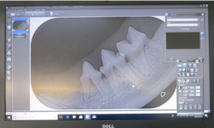 From the examination of the tooth and the x-rays that we have performed, the vet can see that tooth 409 (the right, mandibular molar) has established periodontal disease, causing loss of soft tissue and bone support, especially affecting the bottom of the root. She can also see that tooth 407 (another right, lower molar) has a “type 2 tooth resorption lesion” which has progressed to “stage 3” severity (deep dental enamel loss that extends into the “pulp cavity”, or nerve). Neither of these dental diseases are reversible and the damage will be causing pain, so both of these teeth need to be extracted!
12:35pm
Having teeth removed is painful. As well as a lovely concoction of a pre-med including pre-emptive analgesia, the vet also now administers a local anaesthetic block to the mandibular nerve, which completely numbs the area around the teeth that are to be extracted. The local anaesthetic will last for 6-8 hours, so will carry on providing pain relief for several hours after surgery.
12:45pm
Once the local anaesthetic has had 10-15 minutes to reach full effect, the vet can begin the delicate, skilled procedure of extracting the teeth. Serious trauma and injury can be caused to the patient if this is not performed by a qualified, competent and experienced Veterinary Surgeon, and it should be noted that tooth extractions are never to be carried out by anyone other than a vet. Our vets usually wear loupes (like a jeweller’s magnifying glasses) as cats have very small teeth, with slender, brittle roots, and the specific instruments and suture material used for dentistry are very fine.
The vet uses a surgical flap technique to open the gum, exposing the bone around the tooth, to reveal root surfaces and allow a good repair after extracting the necessary teeth. A small amount of bone is removed from around the tooth roots to allow the vet to see the roots to be extracted. Teeth with more than one root are divided using a high speed, water-cooled drill. Tiny sharp instruments, precise technique and patience are need to stretch the periodontal ligaments and finally remove the tooth root. After extraction any rough bone is smoothed and the tooth socket is cleaned of any debris.
Once the teeth are successfully removed, post extraction radiographs are performed to ensure that all fragments of the tooth have been removed. The extraction sites are then flushed with saline and sutured (stitched up) with a dissolvable suture, to promote fast healing, and prevent any food or debris from becoming trapped in the hole where the tooth was, which can cause pain and infection.
1:05pm
The vet now polishes the remaining teeth, which helps to smooth the tooth surface, making it harder for plaque to accumulate. The mouth is then carefully rinsed out, and an oral mouth wash rinse applied, before the throat pack is removed.
1:10pm
McClane is then allowed to wake up quietly and slowly from his anaesthetic, whilst remaining cuddled up in the warm Bair Hugger blanket and we give him a nice groom (we imagine grooming yourself with a sore mouth is not very nice!). When ready, he is “extubated” (his endotracheal breathing tube is removed) and allowed to breathe oxygen and room air through a face mask.
1:20pm
 Once McClane is fully awake, and all monitoring and anaesthetic equipment has been removed, he is taken back to a hospital pod, temporarily separated from his brother, to recover. We monitor him closely whilst he is in recovery – this is one of the most crucial periods for post-anaesthetic complications.
1:30pm
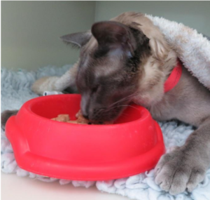
McClane is now ready for his belated breakfast, which he eats very enthusiastically (and with surprising ease, despite his mouth being a little numb after the local anaesthetic block).
1:40pm
McClane continues to recover under close supervision, whilst Mojo is prepped ready for his own dentistry procedure. Despite having significant gingivitis (inflamed gums) present throughout his mouth, Mojo’s teeth only showed early or mild periodontal disease. With no problems identified in his radiographs, treatment with scaling and polishing above and below the gumline was performed and no extractions were needed.
Both boys had several teeth with probing depths of 1mm, so it will be very important for them to receive home dental care (tooth brushing) to prevent these teeth from requiring extraction in the future.
2:45pm
The brothers McClane and Mojo have been reunited, and are tucking into their second breakfasts of the day. Now that they have both recovered well from their surgeries, their owner is phoned to update them that all had gone very well, and to arrange a discharge appointment for later that day.
6pm
Their owners are here to collect them. The vet meets with the boys’ owners to discuss the day’s surgeries, explaining the findings from the radiographs, and providing verbal and written aftercare instructions. As McClane had multiple extractions, he has been given a bottle of pain relief and anti-inflammatory medication, to take at home for the next few days.
Once their intravenous catheters have been removed, temporary pressure bandages applied, and identification collars removed, the brothers are transported into their carriers, and into Reception, ready to go home.
The vet has asked them to come back in 10-14 days for a recheck appointment, but sooner if they have any concerns.
10-14 days later
The brothers are back for their recheck appointment with the vet. They have recovered very well, and the surgical extraction sites in McClane’s mouth are healing nicely. The vet now takes this opportunity to speak to the owners regarding home dental care, and the options available which may suit the personalities and temperaments of the Tonkinese brothers. Prevention of periodontal disease is aimed at controlling plaque. As you can imagine, some cats will tolerate tooth brushing, whereas others certainly will not, so a plaque-repelling granule or oral mouthwash may be more suitable. McClane and Mojo’s owners will be sent a reminder in 6 months to invite them back for a recheck appointment, to see how their mouths are looking.
As you can see, good quality, thorough feline dentistry is quite involved and this really is just the basics of what we get up to behind the scenes. Where multiple extractions are needed, dentistry can be time consuming and incur significant costs.
Cats are the masters of hiding their problems. Even those cats with severe or painful oral health problems may not show any obvious signs, and rarely stop eating.
This is why it is important to have regular health checks with the vet, who can take an experienced peek inside their mouth, and indicate if there is a problem. Carrying out recommended dental work, in a timely manner is very important to preserve your cat’s teeth and limit the progression of dental disease.
If in doubt, contact us today on 01223 880707 (or contact us here)to make an appointment for your cat to have a dental check!
My cat needs dentistry surgery – but what does that actually involve?
People often worry when their cats are coming in to see us for “a dental”. However, tooth disease is very common as cats get older, and treatment (including surgery) is absolutely necessary to maintain a good quality of life. So, in this blog, we’re going to follow two of our recent patients when they came in for dental care with us!
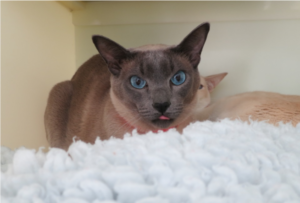
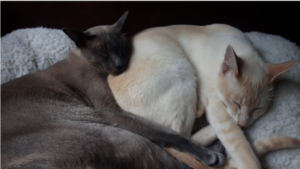 9am – These are Mojo and McClane, who are staying with us for the day to have their dentistry surgeries. They are a very bonded pair, so have come in to the clinic on the same day, to keep each other company. Minimising stress in the clinic is in many cases the key to a successful outcome, so we work very hard to keep our patients as happy and relaxed as possible.
The pair have their admission appointment with vet Mini, who discusses the day’s plan with their owner. She then gives the boys a general health check, and performs any necessary pre-surgery procedures, such as blood pressure measurement and blood sample analysis. Mini then places a paper identification collar on the cats’ necks.
At the end of the admission appointment, Mini asks their owners to read through, and sign, their procedure consent forms. We also ask the owners to record emergency contact details for the day, any belongings left with the cats, and any particular food preferences for their dinner once they have recovered from their anaesthesia. We have a cupboard full of tasty and ‘naughty’ delicacies, too!
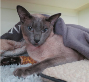
9:40am – The boys are settled into their hospital accommodation. They have their owner’s old jumper with them, along with lots of lovely soft vet-bedding, to help them feel at ease in unfamiliar surroundings. Cats rely heavily on familiar scents to help them feel comfortable and safe, so a well-used blanket, bed or even piece of old clothing should be brought along on the day.
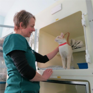
Our hospital accommodation pods contain removable shelves for cats to climb up onto, or hide behind. We also add in litter trays, water bowls, toys, beds, cardboard boxes and blankets or towels for our patients to hide underneath. The hospital has Feliway (a synthetic copy of the pheromone that cats produce, to help create the feelings of safety and familiarity in their own environments) diffused into the room, as well as sprayed onto bedding. We also spray Pet Remedy onto a small towel or blanket, which many cats find very comforting.
As Mojo and McClane are brothers and very bonded, they are housed in the same accommodation pod for the time before their procedures. After their procedures, they will recover separately, but as soon as they wake up, the boys will be reunited once again.
 10am – The Tonkinese twosome are now settled into their accommodation, and are relaxing after the initial stress of travelling to the clinic. Cats do not cope well with stress, and it is very important to allow them ample time to settle and calm before their procedures. This pre-procedure time also allows us to monitor the cats’ vital signs (heart rate, respiratory rate and effort, and blood pressure) whilst they are relaxed (stress can massively affect their vitals) and wait for the test results of any blood or urine analysis taken that morning.
As you can see, they both chose to cuddle up underneath the shelf, with Mojo using McClane to hide behind!
11:30am – All equipment has been set up, and the anaesthetic protocol for each patient individually planned. We are ready to go!
McClane is the first to have his surgery this morning. McClane is cuddled into a carrier to allow him to be safely moved into our prep room, where he will have his pre-medication (pre-med) injection. We tend not to administer injections to patients whilst they are in their hospital accommodation, as we prefer them to see this room as their safe place, where they can rest and recover, without feeling scared.
 Whilst McClane is cuddled up with a blanket in the carrier, the pre-med is gently administered via injection into a muscle, avoiding any unnecessary handling or struggling. A pre-med usually consists of as many as three drugs mixed together which each provide different analgesic (pain relief) and/or sedative effects. Administering a pre-med allows the patients to relax and become a little sleepy before their anaesthetic. This promotes a smoother induction and recovery from the anaesthetic, provide pre-emptive pain relief (thus reducing the pain a patient feels) and allows pre-anaesthesia procedures to be carried out, such as placement of intravenous catheters and administration of oxygen therapy.
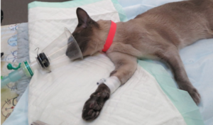
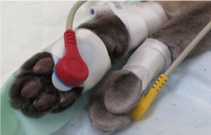
11:45am – After 15-20 minutes, McClane has become a little sleepy, and is happy to be carried to our dentistry table. Whilst he is cuddled up on a warm blanket, an intravenous catheter is placed into a vein in McClane’s front leg. This allows for the administration of anaesthetic drugs, the administration of intravenous fluids, and provides emergency intravenous access.
 When ready, we then administer an intravenous anaesthetic liquid that puts McClane into a state of unconsciousness. We can then pass the endotracheal tube down his trachea (windpipe) which allows us to administer oxygen and anaesthetic gas.
He is then hooked up to various monitoring equipment that provides us with information on his:
– Heart rate, rhythm and electrical output (ECG)
– Pulse rate and peripheral capillary oxygen saturation (SpO2)
– Respiratory rate and rhythm
– Carbon dioxide output (capnograph)
– Temperature
– Blood pressure
As well as all of the fancy equipment, McClane is also monitored continuously, and most importantly, by one of our highly trained Registered veterinary Nurses.
 11:50am – To keep him warm throughout his surgery, McClane is laying on top of a Bair Hugger blanket (warm air is circulated through this), and is covered with plastic wrapping (to trap the warm air) and a thick fleece blanket to keep him nice and cosy. Cats can lose body heat very quickly during anaesthesia, so it is essential to try to keep the process as quick as possible, whilst also providing extra heating support.
We also use several extra pieces of equipment, such as fluid warming devices, and heat and moisture exchange units, which sit on the end of the anaesthetic circuits, to warm and moisten the oxygen that McClane breathes in during his surgery.
McClane is then connected to a syringe driver, which provides carefully calculated measures of intravenous fluid therapy to help maintain his blood pressure throughout the procedure and protect his kidneys. The fluid rate is adjusted throughout the procedure, where necessary.
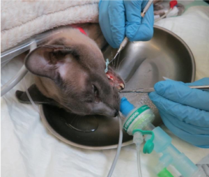
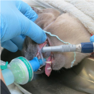
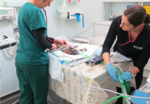
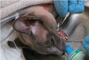 12pm – Once McClane is settled under his anaesthesia, Mini places an absorbent throat pack (cotton wool pad) into the back of his throat, to help absorb any excess fluid, thus further protecting his delicate airways. She then begins to descale his teeth using an ultrasonic descaler. Descaling removes plaque and calculus from above and most importantly below the gum line. A small hand scaler is used to get right under the gum (subgingival). Plaque is the off-white, sticky accumulation on the surface of teeth made up of food particles, bacteria and bacterial products (the furry layer we feel on our teeth in the morning before brushing). Plaque is the main cause of periodontal disease, and turns into Calculus (or tartar/scale) in less than 48 hours after brushing. Calculus can only be removed via descaling, and for several r
easons, cats have to be anaesthetised for this procedure! The mouth is rinsed with water, and then an antiseptic mouthwash after scaling to reduce bacteria before any surgery.
Descaling is more than a cosmetic procedure: it is very important to remove irritants and debris from the teeth and gums in order to treat reversible or early periodontal disease and gingivitis.